“True silence is the rest of the mind, and is to the spirit what sleep is to the body, nourishment and refreshment.”- William Penn
Article authors: Gordon Slater|Tandose Sambo
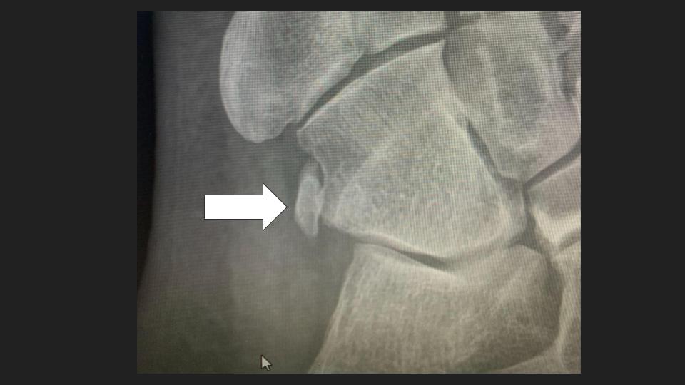
Os peroneum is one of the accessory ossicles of the foot and ankle. The bone is located within the distal peroneus longus tendon and is located lateral to the cuboid[1]. Patients can experience a pain in the midfoot, linked to the os peroneum. The various classifications of this condition include: *Acute Fracture and *Stress Fractures via various root causes. Os peroneum is usually barely noticeable and is detected via medical imaging. A radiograph will be able to identify any mid foot pain. The images provided will be indicative of abnormalities in the foot morphology, and in the location of the os peroneum.
Accessory ossicles such as the os peroneum generally present as asymptomatic, and there are various types of these bones throughout the musculoskeletal system. Orthopaedic surgeons are trained to identify normal locations of the ossicles so that any abnormalities can readily be detected. Various mechanisms for accessory formation are outlined in medical literature. While the os peroneum is generally benign, the root causes of painful os peroneum is often linked to causes such as acute os peroneum fracture, peroneal tendon rupture, and chronic tendinopathy.
Prevalence of the Os peroneum
There are a variety of foot and ankle ossicles and the os peroneum can constitute up to 30% of cases studied. Common foot and ankle injuries include inversion injuries. When patients present with ankle injuries, and experience pain beyond general sprains, os peroneum is often considered. Additionally, if a patient has a peroneus longus tendon tearm the os peroneum can be fractured in the process. Patient symptoms often include pain the ankle, joint instability, swelling of the joint and general tenderness. A physical examination will identify the extent of the condition. Your orthopaedic surgeon will identify the os peroneum as an oval accessory ossicle. Various stages of fragmentation of the os peroneum will also be identified via radiograph.
Treatment of Os peroneum
Treatment of os peroneum has various elements including stabilizing the foot utilization of pain management via NSAIDS and steroid injection. To restore the foot, physical therapy is often administered. In adverse conditions, surgical treatment is an option. Surgical treatments include primary tendon repair, removal of the os peroneum or tendon debridement. The severity of the condition determines the surgical procedure applied.
Reference:
[1] Os Peroneum: https://www.ncbi.nlm.nih.gov/books/NBK538329/