“The human body has been designed to resist an infinite number of changes and attacks brought about by its environment. The secret of good health lies in successful adjustment to changing stresses on the body.” – Harry J. Johnson
What is a Jones Fracture?
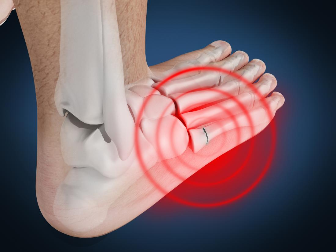
A Jones fracture occurs when the fifth metatarsal often experiences a break between its base and mid region. Like any fracture, patients often do experience pain during a Jones fracture case. Minimally invasive procedures have proven quite effective in the treatment of Jones fractures. Some of the most effective methods of treatment have included the percutaneous fixation of Jones fractures using a 4mm titanium cannulated screw. For patients who this method was applied to, Jones fracture management has proven to be successful. The offices of Dr Gordon Slater are well versed in this procedure, and have conducted extensive research on focus areas such as core tip percutaneous surgical fixation.
What is Core Tip Percutaneous Surgical Fixation?
Core tip percutaneous surgical fixation is a commonly used technique for the management of Jones fracture which otherwise can result in prolonged recovery phase, delayed unions, non unions and risk of refractures. Via previously conducted research, a summary of the treatment of 67 patients who elected to undergo surgical intervention of a Jones fracture between 2007 and 2015 is presented. All patients were fixed with 4mm titanium cannulated screws.
Excessive forces on the feet can result in a Jones fracture. Usually caused by forceful ankle plantarflexion and forefoot adduction, excessive forces placed on the lateral border of the forefoot and metatarsal heads cause the condition. This unique type of fracture includes the articulation of the fourth and fifth metatarsals and is known for its tendency to have delayed healing time. This in turn can result in non-unions and treatment difficulties for clinicians.
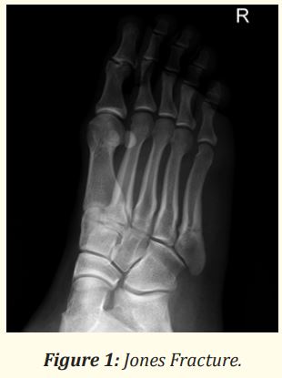
Additional complications such as prolonged healing may be induced due to the development of a watershed area of the blood supply in the metaphyseal-diaphyseal junction. Figure two highlights the different categories of the Jones fractures below:

Jones Fracture Treatments
The treatment procedure for a Jones fracture is directly correlated to the location of the fracture, zone classification and demands or expectations of the patient. Traditionally, the field has facilitated more conservative treatment approaches, but with the advent of technology and the rapid innovative changes, the transition to techniques such as surgical fixation as a treatment of Jones fractures has emerged.
With percutaneous fixation the following are the benefits:
- Union rates can be improved.
- Immobilization times are reduced.
- Refracture rates diminished.
With the classification of Jones fractures, evolving therapies help surgeons tailor their treatment approach based on the specific characteristics of the fracture subtype. As a result, surgeons are able to select the most suitable treatment option for each patient, thereby increasing the likelihood of positive treatment outcomes while simultaneously minimizing the risk of complications. Type I and II fractures can be treated either conservatively with a period of 6 to 8 weeks of non-weight bearing or with surgical intervention. Type III fractures all require surgical intervention due to the severity of the fracture.
Jones Fracture Patient Study
Gordon Slater et al analyzed the treatment outcomes of patients who underwent internal fixation of a Jones fracture using a 4mm titanium cannulated screw by a single surgeon. Sixty seven patients who elected to undergo minimally invasive surgical fixation of Jones fractures were assessed during 2007- 2015. Patients were referred from primary care physicians for surgical opinion, and all surgeries were performed by a single surgeon. The table below identifies the patient categories:

Patients ranged in age from 12 – 72 years old, with a mean age of 38 years. The assessed patient data included:
- Surgery Date
- Etiology of Injury
- Radiographic Findings
- Time from Injury to Surgery
- Time to Radiologic and Clinical Union
- Noted Complications or Secondary Procedures Required
- Return to Pre -injury Activity
- Type of 4mm Cannulated Screw Used
Surgical Procedure: Jones Fracture Minimally Invasive Treatment
The study focused on the assessment of patients who presented within 6-weeks of fracture categorized as acute and treated with screw fixation only. Antibiotic use was also coupled with the therapy.
Surgery Results:
According to the surgery, two types of screws were tested in the study: Smith and Nephew and Integrant screws. Those patients treated with Smith & Nephew 4mm screws (61 patients) were weight bearing at 4 weeks. Those treated with 4mm Integrant screws, (6 patients) were non weight bearing for two weeks and then ambulatory in a cam walker.
Post Surgery Outcomes:
A total of 67 patients (40 females and 27 males), with a mean age of 38 were operated on. Patient results included:
- Successful union achieved in 94% of cases after the primary surgery, with a mean time for clinical union of 8.9 weeks (ranged 4 – 28 weeks).
- Four patients developed infections postoperatively and were treated with a course of oral antibiotics; there were no incidents of deep vein thrombosis or neurological injury.
- No screw breaks or nerve injuries were reported in our study group. Although Asnis cannulated screws were initially selected as the preferred screw, in later cases, Integrant screws were selected, as this new option was more clinically suitable.
- All patients returned to activity after surgical follow ups.
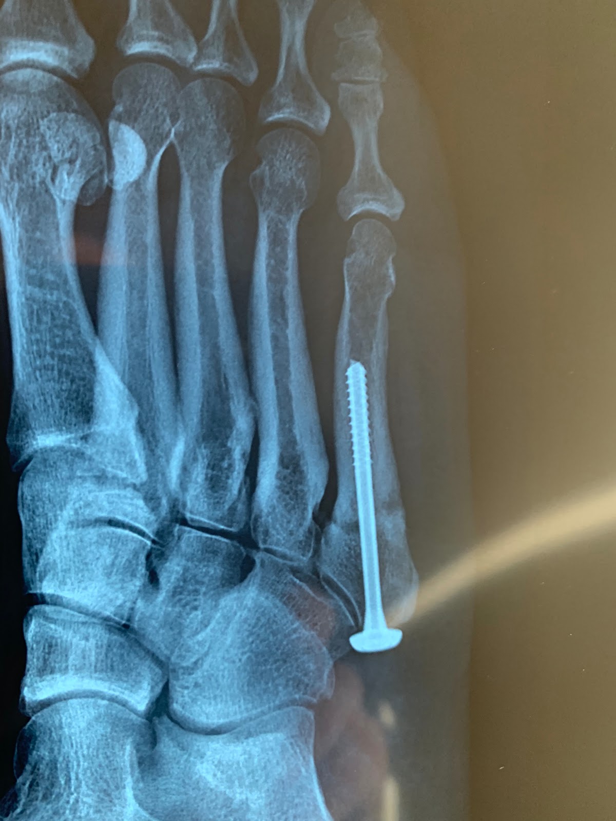
Screw fixation as a therapy has proven its effectiveness as a therapy for the treatment of Jones Fractures. For current patients in our practice, the image below indicates the post op surgical results for a left foot treated with screw fixation:
Jones Fracture Citation:
Gordon L Slater., et al. “Jones Fracture Fixation: Minimally Invasive Management of 67 Cases”. EC Orthopaedics 3.6 (2016): 415- 424. Received: August 10, 2016; Published: August 19, 2016
Distraction Arthroplasty:
