Diabetic Charcot Foot: Causes and Treatments
Article Authors: Gordon Slater| Tandose Sambo
Charcot Foot is defined as a neuropathy induced condition which results in the weakening of the ankle joint, and ultimately the flattening of the feet. Neuropathy patients often can’t feel their extremities due to nerve damage, and ultimately end up deforming the foot as the joint collapses. This condition is quite serious, and several treatments have been proposed, that facilitate the restoration of balance in the body.
Stages of Charcot Foot Development
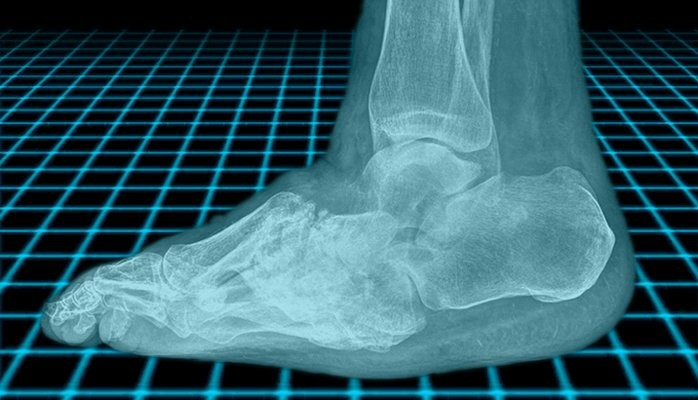
Alternately known as Charcot neuropathic osteoarthropathy (CN), Charcot foot affects the bones, joints and soft tissues in the foot and ankle. Stage one of the condition often involves the inflammation of the joint. Patients often exhibit a condition known as peripheral neuropathy. For diabetic patients, the combination of diabetic complications and neuropathy often leads to the development of Charcot Foot, if the condition is not monitored. For diabetic patients, the factors at play include: diabetes, sensory-motor neuropathy, autonomic neuropathy, trauma, and metabolic abnormalities of bone. The resultant inflammation that is the outcome of these influences can lead to bone degeneration.
As the bone degenerates, the joint ultimately deforms, causing a midfoot collapse that forms the “rocker-bottom” shape that is associated with Charcot Foot. While multiple root causes for the condition may exist, the progression of the patient once the symptoms are onset is as described above.
Potential treatment therapies
Bone and joint destruction is one of the aftermaths of the onset of Charcot Foot. As the condition propagates, the body generates a neurally mediated vascular reflex that leads to increased peripheral blood flow, and accelerates bone resorption. It is anticipated that the counter of this condition – reduction of the peripheral blood flow to the ankle joint, is hoped to stabilize bone health. Studies are currently being conducted that aim to validate this theory. [1] Studies are also being conducted that will identify the root cause of the prolonged, uncontrolled inflammation that patients often experience. The mechanism of this process is currently being elucidated, along with the factors that contribute to neuropathy. Studies have shown that people who have had an acute Charcot foot exhibit retention of vasodilatory reflexes in contrast to diabetic individuals with distal symmetrical neuropathy without CN.[1] Classification of diabetes also has a role to play in the development of Charcot Foot. Patients with type 1 diabetes and type 2 diabetes will have differing tendencies to develop Charcot Foot.
CHARCOT FOOT TREATMENT
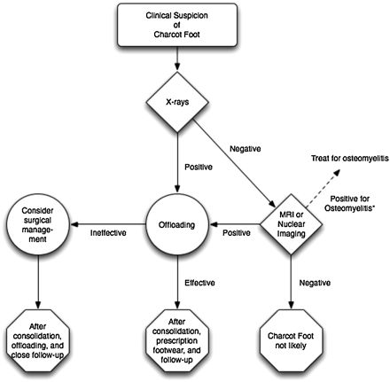
Treatment options for Charcot Foot include surgical reconstruction of the foot. This possibility is the case for most patients, however, there is a pool of patients that are not great candidates for surgery. Under such conditions, the need for a little engineering to restore the feet, will be required. Bracing the feet, is one ideal process for treating patients with Charcot Foot. A decision matrix like the one outlined in the image above, is utilized to determine the path of action. Orthopaedic surgeons do endorse surgical reconstruction of Charcot Foot deformities.
For those patients who are not in the surgery candidate pool, braces will facilitate the desired treatment outcome. The benefits of the braces include the facilitation of mobility while undergoing treatment. As long as the limb can be salvaged, a path of action will be the best one to take. The success rate of bracing is also noteworthy. Orthopaedic surgeons who applied this treatment to their patients have reported good results.
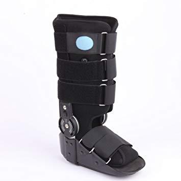
Image Credit: Amazon.com
Considerations prior to Bracing
When you speak to your orthopaedic surgeon, there will be a variety of factors that will be assessed prior to determining whether or not surgery versus braces will be the optimum path for you to take in your Charcot foot treatment. These factors include the inflammation level, the range of motion, and general stability of the foot. For those persons who have been assessed, and the feet have been found to be stable, braces are a good option. Assessing the plane of deformity is another influencer in your health care professional’s decision-making process.
Assessing the various stresses and strains in the feet such as shearing, bending and vertical load is key to optimum restoration. Treatment options for the varying conditions include:
Shearing – Inner soles or orthotics
Bending – Ankle Equinus
Your health is your wealth. If you are diabetic, or suffer from any condition such as Peripheral Artery Disease which affects your cardiovascular system, do ensure that you periodically check with your orthopaedic surgeon to ensure that your feet are not being affected by conditions such as Charcot Foot.
Article Reference:
- The Charcot Foot In Diabetes: https://care.diabetesjournals.org/content/34/9/2123
- Cleveland Clinic: https://my.clevelandclinic.org/health/diseases/15836-charcot-foot
- Stem Cell Applications in Diabetic Charcot Foot and Ankle Reconstructive Surgery: https://www.woundsresearch.com/content/stem-cell-applications-diabetic-charcot-foot-and-ankle-reconstructive-surgery
