Image Credit: All3DP
Introduction
Imagine a world where your orthopaedic treatment is customized to your body. 3-D printing technology is making this possible. With the advances in the economy of the technology, orthopaedic surgeons are now able to visualize the surgery to be performed on a patient, with the 3-D replica of the internal conditions of the patient. Not only that, if necessary part replacement is needed within the body, the bone or tissue needed for repair can also be engineered by your healthcare professional for insertion into your healing site. With better planned surgeries, and body parts customized to your needs, your recovery and healing will be optimized. The future of medicine is truly an amazing concept to behold.
Within medical treatment, there is always an advancement and evolution. The current available treatments all have one thing in common, they are in direct correlation to the available technologies at the time. Before imaging technology, understanding what was taking place within the affected site was difficult. Over the past one hundred years however, the advent of technologies such as computed tomography (CT) , X-rays and magnetic resonance imaging (MRI) are making it possible to see into the body, and facilitate the reconstruction of the site, prior to actual surgical intervention. If the orthopaedic surgeon can see ahead of time, he can plan the surgery accordingly, to ensure the minimal invasive route to be taken for the fastest healing time. 3D printing technology is the bridge between the virtual and the physical world.
The 3-D printing phenomenon is impacting all aspects of our lives. In the industrial realm, the technology is utilized to build in house prototypes for instance. By building the appropriate models for a purpose, the future intention can fully be seen. As a technology that is built on the principles of additive manufacturing, the required orthopaedic model can be built via layering of the desired material into the shape that is required.
The process is a simple one. From the medical image generated via the desired medical image, the segment of bones and ligaments can be reconstructed in such a manner that there is great visibility in the prototype. From the digital twin to the physical twin of the area of interest, various critical organs have been studied and improved by 3-D printing technology. Coupled with a technology such as robotic surgery, healthcare is evolving into an interesting field. Orthopaedics approach to healing patients takes on a different perspective. With access to both the nature of the surgical site, and the tools to heal the problem, the surgery will be a seamless effort.
Orthopaedic procedures often involve the utilization of mechanical interventions to facilitate the healing of the patients. With a pre-prepared model, it will be possible to identify exactly what size of plating, and the appropriate screws will be best for the surgical procedure. The bone and ligament support structures can also be generated for the treatment.
Process of 3-D Printing
As a patient, you should be excited about 3-D printing and the impact that it will have on your healthcare. Think beyond the current condition, and you’ll find that you are in a field of infinite possibilities. For your own home, one day you will be able to 3-D print any items that you may need for your home. If you have a 3-D printer, and you want to create a ceramic vase for instance, you can insert the model of the vase that you want into the printer’s software, along with the relevant materials that you want the vase to be created from, and let the printer produce the final output. The process is similar in the medical realm, and you’ll be able to have your body parts generated in situ, for insertion into your healing site with a custom fit.
For the generation of a 3-D model, there will be a series of mathematical calculations that the system will conduct once it is given the technical specifications from the medical data that is provided from the initial medical images. The printer’s software will take the time to actually assess the image, and identify the contour structures of the image provided. With the appropriate parameters of the image identified, various algorithms will convert these details into a digital format that can be translated into a physical model. The processes involved in this translation include:
- Initial hand drawings of the image, if the medical images aren’t clear enough. On the actual image itself, if the outline of the image is unclear, a manual sketch of the image can be generated and input to the machine.
- Automatic image generation can also be done where necessary. The imaging software can determine via algorithm what the morphology of the image will be.
- In some instances, the manual mode and automatic mode of the image input to the software can be fused together.
Within the computing realm, the input quality is what will determine the quality of the final 3-D printed part. Via computer algorithms, the relevant information is translated downstream until the part is physically generated by the printer.
3-D printing is actually ideal for the prototyping process, and where possible, various iterations of the model can be generated until the desired output is generated. 3D printing is an intricate process. The generated 3-D process is actually virtually sliced into thin layers. Each layer is then added to the layer below it, until the full model is generated. There is a direct correlation between the resolution of the printer, and the outcome generated. With the higher resolution printers is a cost, so in the healthcare realm only the best will do.
Applications of 3-D Printing In the Orthopaedic Realm
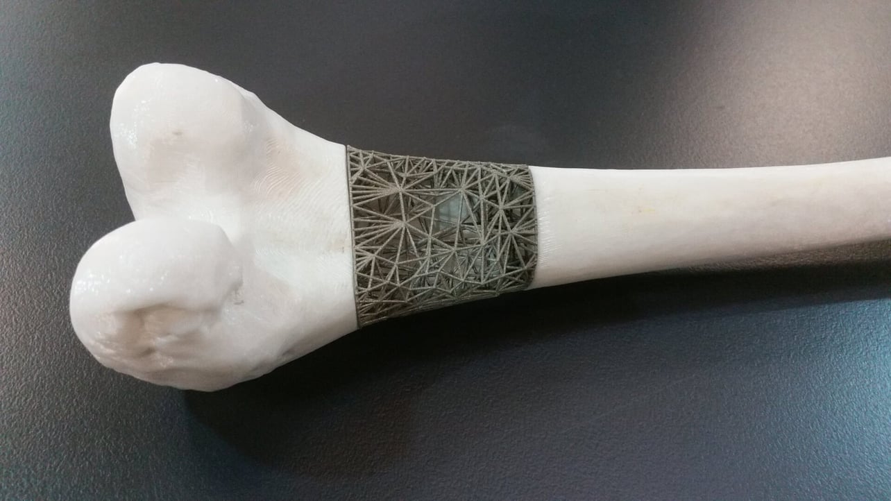
In the orthopaedic realm, the utilization of 3-D printing is becoming quite extensive. From the previous segment in image translation to physical generation, the bone structure images are ones that are well contrasted, and readily deciphered by 3-D printing technology. Because of their clarity, the bone images can be readily segmented and decoded by the system algorithms. Bone structure is ceramic in nature, and for this reason, materials can be manufactured to match the properties desired for this utilization. Technologies such as selective laser sintering are able to print materials such as bones from metal and ceramic powders.
Preliminary models of the clinical case can be generated, in order to give an initial overview of the patient’s case. From the model, pre-surgery practice can be conducted, and utilized in the notes for the actual procedure to be conducted. For the orthopaedic realm, 3-D printing is a welcomed medical innovation. During complicated procedures where there may be conditions such as multiple bone fragmentation, or even in some cases of bone deformities, the 3-D printed replacement can actually be effortlessly facilitated. 3-D printed parts are now being utilized in increasing quantities during the minimally-invasive surgical procedure.
Talk to your orthopaedic surgeon, about whether you feel this procedure might work for you and your current condition. Your health is your wealth.
Reference Article:
3-D Printing: Clinical Applications In Orthopaedics and Traumatology: https://www.ncbi.nlm.nih.gov/pmc/articles/PMC5367547/
