“The higher your energy level, the more efficient your body. The more efficient your body, the better you feel and the more you will use your talent to produce outstanding results.”
-Tony Robbins
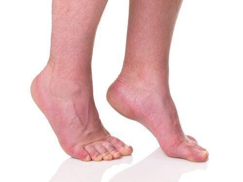
What Is Equinus?
Equinus is a limitation of the upward bending motion of the ankle joint. While most persons can create somewhat of a right angle between their feet and their lower legs, individuals with equinus are unable to do this. As a condition that affects the feet, equinus can occur in either one or both of the feet. If both feet are affected, the severity is different in each foot, as the body adjusts in order to manage the body’s weight.
If left unmanaged, individuals with equinus often find ways to compensate for their limited ankle mobility. Without adequate orthopaedic care, the body will develop additional foot, leg, or back problems. Many persons, for instance, place the majority of their weight on the ball of the foot.
What Causes Equinus?
Equinus has several identified root causes. The first root cause can be attributed to a tightness in the Achilles tendon, or calf muscles. The restriction can be induced either as an inherited trait, or developed during later stages of life. Additional causes of the onset of equinus includes limited mobility of the leg while it’s in a cast, during the process of wearing crutches, or even with the wearing of high heeled shoes. High heels are shoes that women must be careful in wearing because they cause several different foot ailments including hammertoes. Diabetics are also another group that is prone to developing Equinus. During the diabetic process, the condition can affect the Achilles Tendon, and ultimately cause a tightening of the tendon. Underlying neurologic disorders, also associate with diabetes are also responsible for spasms that can affect the ankle joint.
For an externally induced incident of Equinus, if an individual has a fracture of the leg, a fragment of bone can dislodge, and cause a restriction in motion of the ankle. With a variety of root causes that can cause the condition, it is important for the appropriate treatment plan to be put in place that will facilitate restoration of the feet.
Foot Problems Related to Equinus
Depending on how a patient compensates for the inability to bend properly at the ankle, a variety of foot conditions can develop, including[1]:
- Plantar fasciitis (arch/heel pain)
- Calf cramping
- Tendonitis (inflammation in the Achilles tendon)
- Metatarsalgia (pain and/or callusing on the ball of the foot)
- Flatfoot
- Arthritis of the midfoot (middle area of the foot)
- Pressure sores on the ball of the foot or the arch
- Bunions and hammertoes
- Ankle pain
- Shin splints
Diagnosis of Equinus
The best way to classify a case of Equinus, is to consult with an Orthopaedic Specialist. From a medical perspective, equinus is often diagnosed, when the patient is unable to achieve 10° of ankle joint dorsiflexion[2]. What patients often experience before the severe cases of Equinus are the symptoms that are associated with the condition.
There will be a variety of tests that are undergone, in order to diagnose if a patient has equinus. Your foot and ankle surgeon will initially test the range of motion of the ankle. This is done in two states: when the knee is flexed (bent) or straightened. Range of motion tests will identify any tightness in the muscles or tendons.
For an internal view of the ankle, X-rays will also be done. From the aforementioned introduction, there may be some neurologic tests that will be done, in order to ensure that the nervous system is operating as it should.
Nonsurgical Treatment of Equinus
Surgical treatment of any orthopaedic condition, is often an action of last resort. In the initial stages of Equinus, there are a series of actions that your orthopaedic surgeon will prescribe in order to keep your Equinus under control. These include[1]:
- Night splint. The foot may be placed in a splint at night to keep it in a position that helps reduce tightness of the calf muscle.
- Heel lifts. Placing heel lifts inside the shoes or wearing shoes with a moderate heel takes stress off the Achilles tendon when walking and may reduce symptoms.
- Arch supports or Orthotic devices. Custom orthotic devices that fit into the shoe are often prescribed to keep weight distributed properly and to help control muscle/tendon imbalance.
- Physical therapy. To help remedy muscle tightness, exercises that stretch the calf muscle(s) are recommended.
From medical research: “Treatment for equinus should be aimed at increasing ankle joint dorsiflexion to facilitate normal gait mechanics. In most cases, nonspastic forms of equinus tend to be more treatable and easily corrected than the spastic neuro-induced forms of equinus. A variety of conservative measures can be employed, including stretching, bracing, and orthotic devices.
The merits of manual stretching for the gastrocnemius muscle have been a topic of debate, but studies have demonstrated that favorable results are possible when stretching is done correctly even for short time periods. Grady and Saxena found that manual stretching for five minutes per day over six months increased dorsiflexion by an average of 2.7°.23,33 Similarly, Macklin and colleagues were able to elicit excellent results in a group of runners with equinus contractures. Their results produced an average increase in ankle joint dorsiflexion from 5° to 16° following an eight-week stretching program.”[2]
When Is Surgery Needed for Equinus?
If there is an instance of severe Equinus, surgery may be the only option that will ensure an adequate recovery. If there is a bone fragment or a tight tendon, it will be important for surgery to be done. As an inherited trait, the calf muscle tightness is one that often manifests later in life, and surgical treatments can help to alleviate this condition. The surgical path that is decided by your orthopaedic surgeon will be dependent on the case. There will be a series of decision matrices involved, such as the utilization of the Silfverskiöld test to identify your Equinus classification. Depending on the classification, the individual’s surgery will involve either a gastrocnemius recession or an alternate surgical path. With this procedure, there is a noted increase in ankle joint dorsiflexion (range of rotation) of 18.3°.
With such a positive gain to be achieved from surgery, persons with equinus can feel comforted that they will be able to achieve an improvement in their mobility with either non-surgical or surgical procedures.
References:
